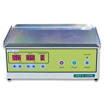
Other Applications using Electrical Cell Fusion
APPLICATIONS
ECFG21 Liposome/droplet Fusion
Timing controllable electrofusion device for aqueous droplet-based microreactors

Fig. 1. Electrofusion of droplets in the fusion chamber.
(a) Droplets formed upstream enter the fusion chamber.
(b) Due to the widening of the chamber, the droplets slow down and make contact when it enters the fusion chamber.
(c) Upon the application of an electric field (50V, 750m gap, Pulse width: 10s, Interval: 0.2sec, 5 times) the contacting droplets coalesce.
(d) Photo showing two coalesced droplets. *The round particle near the top electrode was an air bubble.
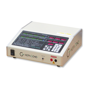
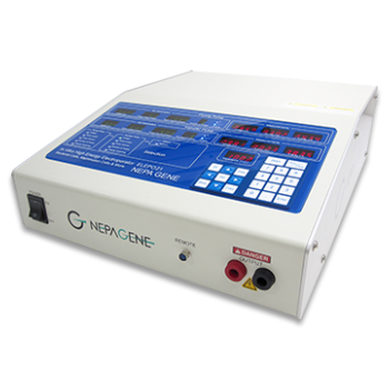
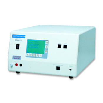
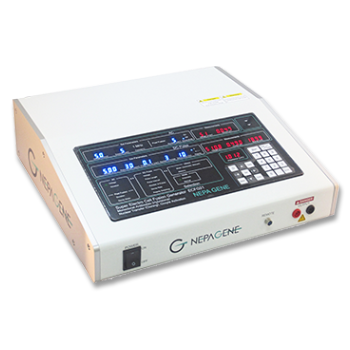
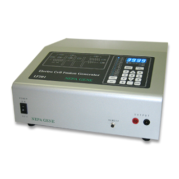
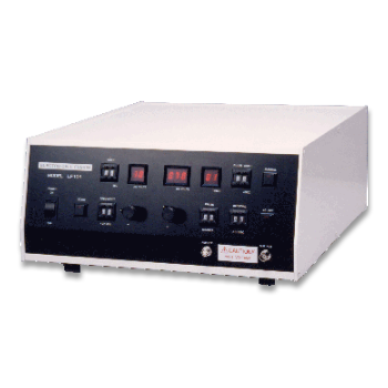
Electric pulses were applied with an Electro Cell Fusion Unit (LF101, NEPA GENE).

Fig. 2. High speed camera images of the fusion process.
This fusion process is almost instantaneous. The two droplets combined into one single ”peanut-shaped” droplet within about 1ms. It took about another 5ms for the droplet to adopt a spherical shape under the effect of surface tension. Throughout the fusion process, the darker colored blue ink droplet (leftmost) was distinctly separated from the lighter colored water droplet (rightmost).
Wei-Heong Tan and Shoji Takeuchi, CIRMM/IIS, Institute of Industrial Science, University of Tokyo
* Lab on a chip, Volume 6, Issue 6, Pages 757-763, June 2006
PUBLICATIONS
- Giant_Unilamellar_Vesicles(GUVs)
Electrofusion Device With High-Aspect-Ratio Electrodes for the Controlled Fusion of Lipid Vesicles
T. Okita, M. Tsugane, K. Kato, K. Shinohara and H. Suzuki.
Journal of Microelectromechanical Systems, vol. 34, no. 2, pp. 174-183, April 2025.
- Giant_Unilamellar_Vesicles(GUVs)
Ejection of Large Particulate Materials from Giant Unilamellar Vesicles Induced by Electropulsation
Katsuta S, Okano T, Koiwai K, Suzuki H.
Langmuir. 2019 Oct 8;35(40):13196-13204.
- Giant_unilamellar_vesicles(GUVs)
A Closed System for Pico-Liter Order Substance Transport from a Giant Liposome to a Cell
Miyakawa S, Uesugi K, Morishima K.
Micromachines (Basel) . 2018 Jul 2;9(7):331.
- Giant_Unilamellar_Vesicles(GUVs)
Introducing Micrometer-Sized Artificial Objects into Live Cells: A Method for Cell–Giant Unilamellar Vesicle Electrofusion
Saito AC, Ogura T, Fujiwara K, Murata S, Nomura SM.
PLoS One . 2014 Sep 17;9(9):e106853.
Electroporation
■ Cell Cultures
- Primary Cell Cultures
- Stem Cells
- Organoids
- Cell Lines
- Cells in Adherence
■ In Vivo Mice/Rats
- Zygotes In Vitro (TAKE method)
- Zygotes In Oviduct (i-GONAD method)
- Embryos In Utero
- Ex Utero Embryos In Vitro
- Brain
- Retina / Cornea / Spinal Cord / Sciatic Nerve
- Lung / Spleen / Liver / Stomach/ Kidney / Intestine
- Pancreas / Islets of langerhans
- Testis / Ovary / Prostate / Gonad / Uterus
- Muscle / Skin / Joint / Cartilage / Tumor / Others
■ In Vivo Other Animals
- Bovine/Porcine/Other Animal Zygotes
- Hamster Zygotes in Oviduct (i-GONAD method)
- Monkey Skin
- Chicken (In Ovo・Others)
- Zebrafish & Other Fishes
- Insects・Others
■ Plant Cells & Algae
- Plant Cells
- Algae
■ Exosomes
- Exosomes
■ Bacteria, Yeast, Fungi
- E. coli/Bacterial Cells
- Yeasts/Fungi
- Bacterial cells/Yeasts/Fungi (NEPA Porator)
Drug Delivery and Transfection
■ Ultrasound Transfection and Drug Delivery (Sonoporation/Fus)
- Brain
- Liver/Skin/Other Applications
- Heart
- Cell Culture
- Lung
- Muscle
Electro Cell Fusion
■ Hybridoma Production
- Monoclonal antibodies, etc
■ Oocyte Activation
- Electrical stimulation before/after Intracytoplasmic sperm injection (ICSI)
■ Somatic cell nuclear transfer (SCNT)・ Oocytes Nuclear Transfer
- Animal cloning
■ Tetraploid Embryos Production
- 2 Cell Embryos (Tetraploid)
■ Other Applications
- Liposome・Protoplast・Yeast, etc.
Fluorescence Quenching / in situ Hybridization Chain Reaction
■ Autofluorescence Quenching
- Mammalian Tissue Sections
- Fish Tissue Sections
- Amphibia Tissue Sections
- Avian Tissue Sections
- Plant Tissue Sections
- Chordate Tissue Sections
■ in situ HCR
- Detection of Target mRNA
Single-Cell/Micro-Particle Transfer
■ マイクロピック&プレースシステム
- Picking and placing micro targets
■ Micro targets
- Animal cells
■ Micro liquid
- Plant cells
Cell Freezing
■ Cell Therapy
- Stem cells, primary cells, and more
■ Animal Husbandry
- Sperm, embryos, tissues, and more

