


Transfection into Chick Embryos in Ovo/Others by Electroporation
APPLICATIONS
Misexpression of the gene of interest by in ovo electroporation

- Injection of the plasmid solution colored by Fast Green into the central canal.
- A pair of electrodes are put on the vitelline membrane overlying the embryos, and a 25V, 50 ms rectangular pulse was charged 4 times.
- We can monitor the transfection efficiency by electroporating GFP expression vector.
- Strong GFP fluorescence could be detected even 48 hours after electroporation.
- Right side of the embryo is transfected, hence the left side could be served as the control. (Dorsal view)
Mechanisms of brain regionalization and neural circuit formation have been studied by misexpression of transcription factors (En1/2, Pax2/5/6, Otx2, Gbx2), secreted factors (Fgf, Shh, semaphoring), signal transduction molecule (Ras, Sprouty2) and receptors (neuropilin).
Harukazu Nakamura and Yuji Watanabe, Graduate School of Life Sciences, and Institute of Development, Aging and Cancer, Tohoku University
*Development Growth & Differentiation, Volume 42, Issue 3, Page 199-201, June 2000
Knock-down by transfection of shRNA expression vector by in ovo electroporation

- shRNA expression vector is electroporated as shown above.
- Select target DNA sequence of 19 to 21 mer. Sense and antisense sequence were linked to a nucleotide spacer as a loop and put into expression vector that is driven by U6 or H1 promoter. Commercially available expression vector that dreives expression by mouse U6 promoter is effective in chick embryos.
- After transcription, hairpin is digested to form siRNA, and siRNA forms RISK (RNA-induced silencing complex) to dgrade the target mRNA.
24 hours after electroporation

- Transfection is monitored by co-electroporated GFP fluorescence.
- Application of siRNA against En2 by electroporation shows degradation of En2 mRNA.
- Control side
Harukazu Nakamura and Tatsuya Katahira, Graduate School of Life Sciences, and Institute of Development, Aging and Cancer, Tohoku University
*Mechanisms of Development, Volume 121, Issue 9, Pages 1137-1143, September 2004
*Development Growth & Differentiation, Volume 45, Issue 4, Pages 361-367, August 2003
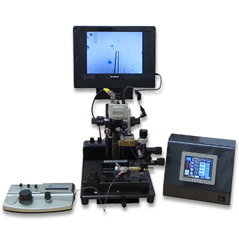
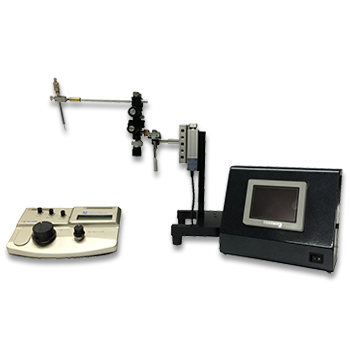
Electroporation for early chick embryos using New's culture (gastrula)

- Schema illustrating the system.
- Hamburger and Hamilton (HH) stage 4 chick embryo with DNA solution injected to the prospective neural plate region. DNA solution was colored with a FastGreen dye.hn: Hensen’s node, ps: primitive streak, ao: area opaca, ap: area pellucida
- Schema illustrating the location of DNA solution between the vitelline membrane and the ectoderm.
Experimental Procedure
- Dissect an embryo using a paper ring (dried filter paper) out of the egg.
- Rinse gently to remove excess yolk, and place the embryo onto the platform of the cathode chamber.
- Inject DNA solution into the space between the ectoderm and vitelline membrane using a glass pipette (for introduction to the ectoderm derivatives).
- Adjust the target site right onto the cathode, and set the anode above it. Set the electrode distance at 5mm.
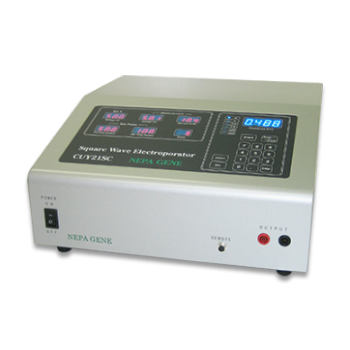
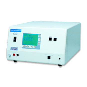
- Deliver electric pulses (10V, pulse on 50ms, pulse of 100ms, 5 pulses) using a CUY21 electroporator.
- Place the specimen on an agarose-albumin plate, and incubate at 39C
Transgene expression after the electroporation
GFP expression becomes detectable by HH6 (about 5 hours after electroporation) in the neural plate, which spreads throughout the central nervous system at HH17 (Scale bar: 1mm).

- GFP gene was introduced to the prospective neural plate at HH4, and the embryo was cultured for about 34 hours (HH17 equivalent).
- GFP expression was occasionally monitored under a fluorescent dissecting microscope.
- GFP fluorescence was detectable as early as 3 hours after electroporation at the central nervous system and head ectoderm.
- More than 80% of cells within the transfected area express GFP.
Gene transfection for tissues other than the CNS

Based on the fate map of the chick epiblast, transgenes can be introduced to the somites (A, E), hematopoietic system (B, F), notochord (C, G), or lateral plate mesoderm (D, H).
E-H are the high power views of A-D, respectively.
Kenji Shimamura, Division of Morphogenesis, Institute of Molecular Embryology and Genetics, Kumamoto University
PUBLICATIONS
- In_ovo
- Anterior_primitive_streak
Mechanical Coupling Coordinates the Co-elongation of Axial and Paraxial Tissues in Avian Embryos
Xiong F, Ma W, Bénazéraf B, Mahadevan L, Pourquié O.
Dev Cell. 2020 Nov 9;55(3):354-366.e5.
- In_ovo
- Ciliary ganglion(CG)
Analysis of Neuro-Neuronal Synapses Using Embryonic Chick Ciliary Ganglion via Single-Axon Tracing, Electrophysiology, and Optogenetic Techniques
Egawa R, Yawo H.
Curr Protoc Neurosci. 2019 Apr;87(1):e64.
- In_ovo
- Neural_plate
- Neural_tube
Method for electroporation for the early chick embryo
Hatakeyama J, Shimamura K.
Dev Growth Differ. 2008 Aug;50(6):449-52.
- Ex_ovo
- Neural_plate
- Neural_tube
Apical Accumulation of Rho in the Neural Plate Is Important for Neural Plate Cell Shape Change and Neural Tube Formation
Kinoshita N, Sasai N, Misaki K, Yonemura S.
Mol Biol Cell. 2008 May;19(5):2289-99.
- Ex_ovo
- Anterior_primitive_streak
Dual mode of paraxial mesoderm formation during chick gastrulation
Iimura T, Yang X, Weijer CJ, Pourquié O.
PNAS, Volume 104, Number 8, Pages 2744-2749, 20 February 2007
- In_ovo
- Neural_tube
Gain- and loss-of-function in chick embryos by electroporation
Nakamura H, Katahira T, Sato T, Watanabe Y, Funahashi J.
Mech Dev. 2004 Sep;121(9):1137-43.
- In_ovo
- Neural_tube
- Brain
RGM and its receptor neogenin regulate neuronal survival
Matsunaga E, Tauszig-Delamasure S, Monnier PP, Mueller BK, Strittmatter SM, Mehlen P, Chédotal A.
Nat Cell Biol. 2004 Aug;6(8):749-55.
- Ex_ovo
- Somites
A primer on using in ovo electroporation to analyze gene function
Krull CE.
Dev Dyn. 2004 Mar;229(3):433-9.
- In_ovo
- Primitive_cardiac_tube
- Ex_ovo
- Cardiac_explants
Dual roles of Sema6D in cardiac morphogenesis through region-specific association of its receptor, Plexin-A1, with off-track and vascular endothelial growth factor receptor type 2
Toyofuku T, Zhang H, Kumanogoh A, Takegahara N, Suto F, Kamei J, Aoki K, Yabuki M, Hori M, Fujisawa H, Kikutani H.
Genes Dev . 2004 Feb 15;18(4):435-47.
- In_ovo
- Neural_fold
- Neural_tube
- Ex_ovo
- Neural_tube_explants
Multiple roles of Sox2, an HMG-box transcription factor in avian neural crest development
Wakamatsu Y, Endo Y, Osumi N, Weston JA.
Dev Dyn. 2004 Jan;229(1):74-86.
- In_ovo
- Neural_tube
Gene silencing in chick embryos with a vector-based small interfering RNA system
Katahira T, Nakamura H.
Dev Growth Differ. 2003 Aug;45(4):361-7.
- In_ovo
- Prospective_esophago_tracheobronchial_region
Tbx4-Fgf10 system controls lung bud formation during chicken embryonic development
Sakiyama J, Yamagishi A, Kuroiwa A.
Development. 2003 Apr;130(7):1225-34.
- In_ovo
- Neural_tube
Inductive signal and tissue responsiveness defining the tectum and the cerebellum
Sato T, Araki I, Nakamura H.
Development 2001 Jul;128(13):2461-9.
Electroporation
■ Cell Cultures
- Primary Cell Cultures
- Stem Cells
- Organoids
- Cell Lines
- Cells in Adherence
■ In Vivo Mice/Rats
- Zygotes In Vitro (TAKE method)
- Zygotes In Oviduct (i-GONAD method)
- Embryos In Utero
- Ex Utero Embryos In Vitro
- Brain
- Retina / Cornea / Spinal Cord / Sciatic Nerve
- Lung / Spleen / Liver / Stomach/ Kidney / Intestine
- Pancreas / Islets of langerhans
- Testis / Ovary / Prostate / Gonad / Uterus
- Muscle / Skin / Joint / Cartilage / Tumor / Others
■ In Vivo Other Animals
- Bovine/Porcine/Other Animal Zygotes
- Hamster Zygotes in Oviduct (i-GONAD method)
- Monkey Skin
- Chicken (In Ovo・Others)
- Zebrafish & Other Fishes
- Amphibian・Insects・Others
■ Plant Cells & Algae
- Plant Cells
- Algae
■ Exosomes
- Exosomes
■ Bacteria, Yeast, Fungi
- E. coli/Bacterial Cells
- Yeasts/Fungi
- Bacterial cells/Yeasts/Fungi (NEPA Porator)
Drug Delivery and Transfection
■ Transfection/Microinjection
- Plant cells & Algae
■ Ultrasound Transfection and Drug Delivery (Sonoporation/Fus)
- Brain
- Liver/Skin/Other Applications
- Heart
- Cell Culture
- Lung
- Muscle
Electro Cell Fusion
■ Hybridoma Production
- Monoclonal antibodies, etc
■ Oocyte Activation
- Electrical stimulation before/after Intracytoplasmic sperm injection (ICSI)
■ Somatic cell nuclear transfer (SCNT)・ Oocytes Nuclear Transfer
- Animal cloning
■ Tetraploid Embryos Production
- 2 Cell Embryos (Tetraploid)
■ Other Applications
- Liposome・Protoplast・Yeast, etc.
Fluorescence Quenching / in situ Hybridization Chain Reaction
■ Autofluorescence Quenching
- Mammalian Tissue Sections
- Fish Tissue Sections
- Amphibia Tissue Sections
- Avian Tissue Sections
- Plant/Algae Tissue Sections
- Chordate Tissue Sections
■ in situ HCR
- Detection of Target mRNA
Single-Cell/Micro-Particle Transfer
■ Micro targets
- Animal cells
■ Micro liquid
- Plant cells
Cell Freezing
■ Cell Therapy
- Immune cells, stem cells, and more
■ Reproductive Medicine & Animal Husbandry
- Sperm, embryos, tissues, and more