
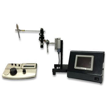
Transfection into Mouse/Rat Testis, Ovary, Prostate, Gonad, and Uterus by Electroporation
APPLICATIONS
Electroporated Transgene-Rescued Spermatogenesis in Infertile Mutant Mice with a Sertoli Cell Defect
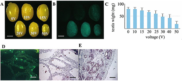
Stereomicroscopic views of transfected testes charged with various voltages and observed under visible (A) or excitation (B) light after 5 wk.
Voltage is indicated on each testis (A).
Loss of testicular weight 5 wk after electric charge in 12-day-old testes (C).
All values are means ![]() SD (measured number of testes charged with 0, 10, 15, 20, 25, 30, 40, and 50 V are 4, 4, 4, 12, 12, 15, 10, and 12, respectively).
SD (measured number of testes charged with 0, 10, 15, 20, 25, 30, 40, and 50 V are 4, 4, 4, 12, 12, 15, 10, and 12, respectively).
Cross-sections of a testis at 5 wk after being charged with 50 V, observed with fluorescence microscope under excitation light (D, left), followed by counterstaining of the same section with hematoxylin (D, right) or another section just stained with hematoxylin (E).
In higher voltage charged testes, luminal enlargement of seminiferous tubules (D) or complete degeneration of seminiferous tubules under the capsule (D, left) was observed. In these testes, many fluorescence-positive Sertoli cells were identified easily (E, right), like in 20V charged testes (Day 35).
Bars = 2mm (A, B) or 100μm (D, E).
Kentaro Yomogida, School of Human Environmental Sciences, Mukogawa Women’s University
*BIOLOGY of REPRODUCTION, Volume 67, Issue 3, Pages 712-717, September 2002
PUBLICATIONS
Electroporation
■ Cell Cultures
- Primary Cell Cultures
- Stem Cells
- Organoids
- Cell Lines
- Cells in Adherence
■ In Vivo Mice/Rats
- Zygotes In Vitro (TAKE method)
- Zygotes In Oviduct (i-GONAD method)
- Embryos In Utero
- Ex Utero Embryos In Vitro
- Brain
- Retina / Cornea / Spinal Cord / Sciatic Nerve
- Lung / Spleen / Liver / Stomach/ Kidney / Intestine
- Pancreas / Islets of langerhans
- Testis / Ovary / Prostate / Gonad / Uterus
- Muscle / Skin / Joint / Cartilage / Tumor / Others
■ In Vivo Other Animals
- Bovine/Porcine/Other Animal Zygotes
- Hamster Zygotes in Oviduct (i-GONAD method)
- Monkey Skin
- Chicken (In Ovo・Others)
- Zebrafish & Other Fishes
- Insects・Others
■ Plant Cells & Algae
- Plant Cells
- Algae
■ Exosomes
- Exosomes
■ Bacteria, Yeast, Fungi
- E. coli/Bacterial Cells
- Yeasts/Fungi
- Bacterial cells/Yeasts/Fungi (NEPA Porator)
Drug Delivery and Transfection
■ Ultrasound Transfection and Drug Delivery (Sonoporation/Fus)
- Brain
- Liver/Skin/Other Applications
- Heart
- Cell Culture
- Lung
- Muscle
Electro Cell Fusion
■ Hybridoma Production
- Monoclonal antibodies, etc
■ Oocyte Activation
- Electrical stimulation before/after Intracytoplasmic sperm injection (ICSI)
■ Somatic cell nuclear transfer (SCNT)・ Oocytes Nuclear Transfer
- Animal cloning
■ Tetraploid Embryos Production
- 2 Cell Embryos (Tetraploid)
■ Other Applications
- Liposome・Protoplast・Yeast, etc.
Fluorescent Staining
■ Autofluorescence Quenching
- Mammalian Tissue Sections
- Fish Tissue Sections
- Amphibia Tissue Sections
- Avian Tissue Sections
- Plant Tissue Sections
- Chordate Tissue Sections
■ in situ Hybridization Chain Reaction (HCR)
- Fluorescence Detection of Target mRNA
Single-Cell/Micro-Particle Transfer
■ Micro targets
- Animal cells
■ Micro liquid
- Plant cells
Cell Freezing
■ Cell Therapy
- Stem cells, primary cells, and more
■ Animal Husbandry
- Sperm, embryos, tissues, and more