
Transfection into iPS Cells / ES Cells / Other Stem Cells by Electroporation
APPLICATIONS
Transfection Results with Nepa Gene Electroporation System
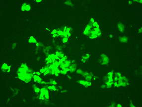
Human iPS Cells
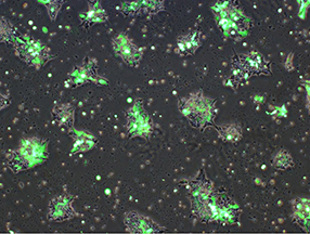
3 days after electroporation
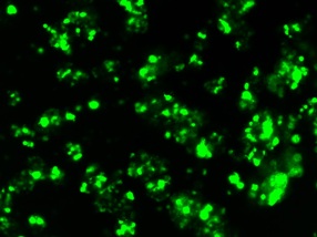
Human iPS Cells
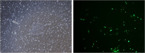
2 days after electroporation
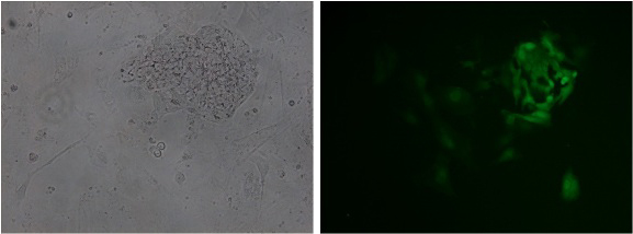
7 days after electroporation
GFP is still expressed in the iPS cell colonies after cell passaging.
Mouse ES Cells
Viability: 74%
Transfection Efficiency: 88%
Mouse Neurospheres
Viability: 90%
Transfection Efficiency: 75%
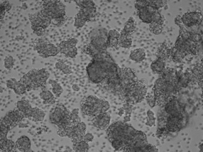
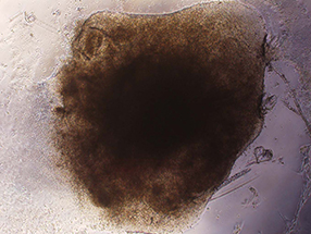
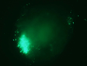
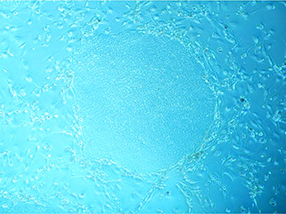
Embryoid Body in Adherence from human iPS Cells
Generation of Induced Pluripotent Stem Cells (iPSCs)
Data of disease-specific iPSC Generation (disease: LQT)
Transfection of multiple episomal plasmids into B cells
iPSC colony one month after electroporation. (5X objective)
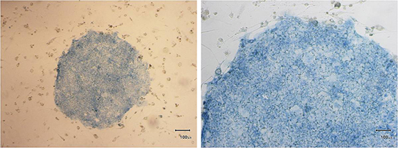
Alkaline phosphatase stain (Left: 4X objective Right: 10X objective)
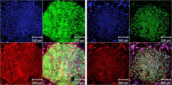
Good expressions of stem cell markers TRA1-60 and OCT4.
(left green: TRA1-60, right green: OCT4, blue: DAPI, red: Phalloidin)
Courtesy of Dr. Toshio Nakanishi laboratory, Department of Pediatric Cardiology, Tokyo Women’s Medical University, Japan
Evaluation of CRISPR activity of human iPS cells with NEPA21 transfection

- Optimization of transfection conditions using the NEPA21 super electroporator (Nepa Gene Co., Ltd.) in human iPS cell lines. Voltage and pulse width of Poring Pulse were examined. EGFP expression plasmids were transfected and the percentage of positive cells was analyzed using the LSRFortessa Cell Analyzer (BD).
- Schematic of transfection procedure.
- Cas9 expression vector pHL-EF1a-SphcCas9-iP-A (Addgene ID: 60599) and sgRNA expression vector pHL-H1-ccdB-mEF1a-RiH (Addgene ID: 60601) were electroporated into iPS cells and then the endogenous dystrophin gene cleavage activity was evaluated by the T7EI assay. The intensity of the cleaved bands (◀ red arrows) was detected with a TapeStation (Agilent) and the values are shown below the gel electrogram images.
- The same genomic DNA samples as in (C) were analyzed by RFLP with restriction enzyme XcmI (50-CCANNNNN^NNNTGG-30). The intensity of the uncut bands is indicated by ◀ red arrows. Because gRNA5 cleaves far from the XcmI site, no cleavage activity was detected in the RFLP assay.
Courtesy of Dr. HongMei Li and Dr. Akitsu Hotta, Center for iPS Cell Research and Application (CiRA), Kyoto University
Transfection Data: iPS Cells / ES Cells / Other Stem Cells
See the cell images by clicking the cell names.
V: Viability, TE: Transfection Efficiency.
| Cells name | V | TE | Cells name | V | TE | |
| Human iPS Cells (201B7) | 86% | 70% | Human iPS Cells | 94% | 80% | |
| Human iPS Cells | Human iPS Cells | |||||
| Human iPS Cells (201B7) | 85% | 94% | Human iPS Cells (201B7) | |||
| Human iPS Cells | 69% | 80% | Human iPS Cells | 73% | ||
| Human iPS Cells Derived Neural Cells | 93% | 54% | Human ES Cells | |||
| Human ES Cells (H9 p.51)) | 55% | 55% | Human Mesenchymal Stem Cells | 96.2% | 96.7% | |
| Human Mesenchymal Stem cells (Primary) | 78% | 75% | Human Mesenchymal Stem Cells | 70% | 80% | |
| Human Neural Stem Cells | 97% | 95% | Human Neural Stem Cells | 80% | 83% | |
| Human Deciduous Teeth Stem Cells (SHED) | 90% | 92% | Human Nucleated Cells Including Hematopoietic Stem Cells (Before cell isolation) | 73% | 90% | |
| Human Adipose-derived Stem Cells (ASC P5) | 87% | 65% | ||||
| Mouse iPS Cells | 70% | 50% | Mouse ES Cells | 80% | 75% | |
| Mouse ES Cells | 80% | 68% | Mouse ES Cells | 74% | 88% | |
| Mouse ES cells (129 strain, R1/E) | 80% | 90% | Mouse ES Cells | 70% | 100% | |
| Mouse ES Cells | 80% | 90% | Mouse iPS cell derived Neural Stem Cells | 86% | ||
| Mouse Neural Stem Cells | 90% | 80% | Mouse Neural Stem cells (Primary) | 80% | 60% | |
| Mouse Neurospheres | 90% | 75% | Mouse Neurospheres | |||
| Mouse Trophoblast Stem Cells | 59% | 47% | C3H/10T1/2 Mouse Mesenchymal Stem Cells | 70% | 85% | |
| Mouse Mesenchymal Stem Cells | 99% | 89% | Mouse Hematopoietic Stem Cells (c-Kit positive cells) | 66% | 45% | |
| Rat ES Cells | 70% | 76% | Rat ES Cells | 60% | 80% |
We have a lot of data of iPS/ES/Other stem cells transfection with high efficiency and high viability. Please feel to free to contact us for the latest data.
PUBLICATIONS
- Human_dental_pulp_stem_cells(DPSCs)
Novel Gain-Of-Function Mutation of TRPV4 Associated With Accelerated Chondrogenic Differentiation of Dental Pulp Stem Cells Derived From a Patient With Metatropic Dysplasia
Nonaka K, Han X, Kato H, Sato H, Yamaza H, Hirofuji Y, Masuda K.
Biochem Biophys Rep. 2019 May 17:19:100648.
- Rat_hair_follicle_stem_cells
miR‑339‑5p negatively regulates loureirin A‑induced hair follicle stem cell differentiation by targeting DLX5.
Li X, Wu Y, Xie F, Zhang F, Zhang S, Zhou J, Chen D, Liu A
Mol Med Rep. 2018 Aug;18(2):1279-1286.
- Human_adipose_mesenchymal_stem_cells(hAMSCs)
CRISPR/Cas9-Mediated Knockin Application in Cell Therapy: A Non-viral Procedure for Bystander Treatment of Glioma in Mice.
Meca-Cortés O, Guerra-Rebollo M, Garrido C, Borrós S, Rubio N, Blanco J
Mol Ther Nucleic Acids. 2017 Sep 15:8:395-403.
Electroporation
■ Cell Cultures
- Primary Cell Cultures
- Stem Cells
- Organoids
- Cell Lines
- Cells in Adherence
■ In Vivo Mice/Rats
- Zygotes In Vitro (TAKE method)
- Zygotes In Oviduct (i-GONAD method)
- Embryos In Utero
- Ex Utero Embryos In Vitro
- Brain
- Retina / Cornea / Spinal Cord / Sciatic Nerve
- Lung / Spleen / Liver / Stomach/ Kidney / Intestine
- Pancreas / Islets of langerhans
- Testis / Ovary / Prostate / Gonad / Uterus
- Muscle / Skin / Joint / Cartilage / Tumor / Others
■ In Vivo Other Animals
- Bovine/Porcine/Other Animal Zygotes
- Hamster Zygotes in Oviduct (i-GONAD method)
- Monkey Skin
- Chicken (In Ovo・Others)
- Zebrafish & Other Fishes
- Amphibian・Insects・Others
■ Plant Cells & Algae
- Plant Cells
- Algae
■ Exosomes
- Exosomes
■ Bacteria, Yeast, Fungi
- E. coli/Bacterial Cells
- Yeasts/Fungi
- Bacterial cells/Yeasts/Fungi (NEPA Porator)
Drug Delivery and Transfection
■ Transfection/Microinjection
- Plant cells & Algae
■ Ultrasound Transfection and Drug Delivery (Sonoporation/Fus)
- Brain
- Liver/Skin/Other Applications
- Heart
- Cell Culture
- Lung
- Muscle
Electro Cell Fusion
■ Hybridoma Production
- Monoclonal antibodies, etc
■ Oocyte Activation
- Electrical stimulation before/after Intracytoplasmic sperm injection (ICSI)
■ Somatic cell nuclear transfer (SCNT)・ Oocytes Nuclear Transfer
- Animal cloning
■ Tetraploid Embryos Production
- 2 Cell Embryos (Tetraploid)
■ Other Applications
- Liposome・Protoplast・Yeast, etc.
Fluorescence Quenching / in situ Hybridization Chain Reaction
■ Autofluorescence Quenching
- Mammalian Tissue Sections
- Fish Tissue Sections
- Amphibia Tissue Sections
- Avian Tissue Sections
- Plant Tissue Sections
- Chordate Tissue Sections
■ in situ HCR
- Detection of Target mRNA
Single-Cell/Micro-Particle Transfer
■ Micro targets
- Animal cells
■ Micro liquid
- Plant cells
Cell Freezing
■ Cell Therapy
- Immune cells, stem cells, and more
■ Reproductive Medicine & Animal Husbandry
- Sperm, embryos, tissues, and more